
Advanced Cone Beam CT Imaging in Anchorage
 When you’ve tried multiple approaches, seen several providers, or had imaging that didn’t provide a clear explanation for your symptoms, a closer look may help. Cone beam CT (CBCT) imaging gives our doctors a detailed, three-dimensional view of your skull, upper neck, and jaw, providing precise anatomical measurements to help guide your care.
A Detailed 3D Look at Your Upper Neck
Cone beam computed tomography (CBCT) captures hundreds of thin image slices of your head and neck, which a computer assembles into a detailed 3D model we can view from virtually any angle. Picture slicing an apple thinner and thinner until you can see the skin, the flesh, the core, and finally the seed.
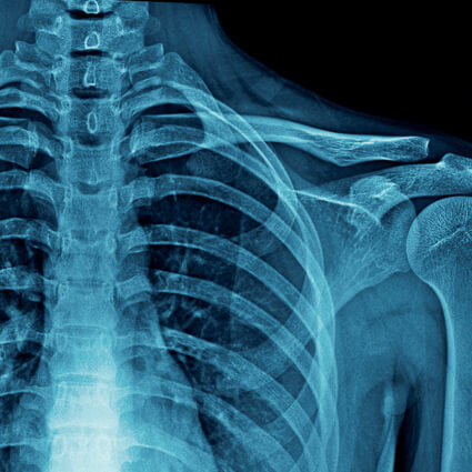
Traditional X-rays flatten three-dimensional anatomy onto a flat image, where structures overlap and positioning differences can affect what we see. CBCT reduces some of these limitations. It allows us to examine the relationship between your skull, your atlas (C1), and your axis (C2), along with degenerative changes, congenital variations, bony changes associated with previous injuries, and TMJ structures that may be difficult to evaluate on standard films.
Who May Benefit From a Closer Look?
The way your upper neck is put together is as unique as your fingerprints. Small anatomical differences can influence how our chiropractors evaluate the relationship between the skull and upper cervical vertebrae.
At Pairmore & Young: Synergy Chiropractic, CBCT may be recommended when a detailed assessment of the upper cervical spine could help guide clinical decisions, particularly for patients considering Atlas Orthogonal care.
Patients who may be evaluated for CBCT imaging include those experiencing:
- Chronic headaches or migraines
- Persistent neck pain and stiffness
- Whiplash and motor vehicle accident injuries
- Neck concerns following a concussion or traumatic brain injury
- Jaw pain and TMJ disorders
- Structural asymmetry and chronic postural problems
- Complex cases that haven’t responded to previous care
CBCT is primarily used to examine bony structures. It does not diagnose concussions or assess brain injuries, but it may provide useful information about associated upper cervical structures when clinically indicated.

From a 30-Second Scan to an Adjustment Planned for You
The scan itself is quick and non-invasive. You simply sit or stand while the scanner rotates around your head and neck for approximately 30 seconds. There are no injections, dyes, or special preparation required.
Our doctors then examine the images in detail, looking closely at the relationship between individual bones. That analysis takes time, so your first adjustment may follow several hours or up to a day later.
For patients flying in from other parts of Alaska or traveling from farther away, we typically arrange imaging in the morning and, when scheduling allows, the first adjustment after lunch.
Detailed Imaging With Radiation Exposure in Mind
For perspective, published estimates for some CBCT examinations range from approximately 0.09 to 0.27 mSv, while the average person in the United States receives roughly 3 mSv annually from natural background radiation. Actual exposure depends on the scan settings and area being examined.
Because any imaging involving radiation deserves careful consideration, our doctors review your needs, examination findings, and imaging history before recommending a scan.
Insurance coverage for CBCT varies. Our team can explain the fees and discuss your payment options before you proceed.

Decades of Experience With Today’s Imaging Technology
Dr. John Pairmore and Dr. Dennis Young bring more than 60 years of combined clinical experience and have worked with thousands of patients experiencing a range of concerns.
Dr. Young has practiced upper cervical chiropractic care for more than 35 years, and he’s particularly enthusiastic about the detail CBCT provides.
For our chiropractors, investing in this technology reflects a longstanding commitment to incorporating advances that may help them better evaluate each patient’s anatomy and plan individualized care. They also participate in ongoing professional education to stay informed about developments in imaging and chiropractic practice.
A Closer Look May Help Guide Your Next Step
If you’ve been told everything looks normal but you’re still dealing with persistent symptoms, a more detailed evaluation of your upper neck may provide additional information to guide your care.
Contact Pairmore & Young: Synergy Chiropractic to discuss whether cone beam CT imaging may be appropriate for you.
